


Osteoarthritis is not an inflammatory disease of the joints, in which there is destructive-dystrophic processes, such as the defeat of the articular cartilage, ligaments and bones, pathological changes in the synovial membrane (or membrane) capsule and the articular (in the joint shell, hermetically closes it is the bonding to the bones in the joint). When osteoarthritis pain is felt in the joint may limit mobility.
Causes of osteoarthritis
Still unknown what is the root cause of osteoarthritis, but there are several factors that worsen the condition of the joint, changing its structure and functioning. The impetus for osteoarthritis can provide:
- trauma (sprains, torn ligaments, fracture, wound, penetrating wound);
- static and dynamic loads associated with professional activity (in athletes, dancers, miners and other persons engaged in heavy physical exertion);
- obesity (there is a constant stress on the joint);
- hypothermia;
- infectious diseases (tuberculosis, chlamydia, influenza);
- circulatory disorders;
- hormonal changes (pregnancy, menopause, menopause);
- of metabolic disorders (osteoporosis (reduced bone density), arthritis (inflammatory joint disease), gout (deposition of uric acid salts in the joints));
- autoimmune diseases (systemic lupus erythematosus (a disease in which the immune system recognizes its own cells as foreign and fights with them), rheumatoid arthritis (the immune system mistakenly invades the healthy joint));
- hemophilia (due to hemophilia frequent bleeding in the joints);
- disorders of the endocrine system (thyroid disease);
- degenerative changes (Perthes disease (aseptic necrotic process in the head of the hip joint), the necrotic process in which the cartilage separates from the bone and moves into the joint cavity, causing necrosis of cancellous bone, resulting in formation of small fractures;
- congenital diseases (connective tissue dysplasia (improper formation of the tissue), hip dysplasia (abnormal development of the joint), congenital abnormalities of the upper and lower limbs);
- old age;
- intoxication (an overdose of drugs, excessive drinking, drugs);
- surgery on the joint;
- sedentary lifestyle;
- heredity;
- beriberi.
The degree of osteoarthritis
The process of the defeat of arthrosis is divided into 4 degrees:
-
The zero degree of osteoarthritis – an obvious morphological changes were observed, but the allusions to the disease is already there. There is a change in the composition of the synovial fluid, so the power supply to the cartilage deteriorates, hence its resistance to the load is reduced. In the morning after awakening or after prolonged rest in a stationary state, there is difficulty in movement of joints, but during the day the stiffness to pass. This condition is called "starting pain". When moving the joint makes a crunching, but the pain during movement do not exist. For the first characterized by pain after a long, hard load on the joint, but the pain disappears immediately after the break.
-
First, the degree of arthrosis, second degree is characterized by destruction of hyaline cartilage. Start morphological changes. X-ray shows the presence of newly formed osteophytes on the edges of the bone surface. The synovial capsule becomes thinner. It is noted narrowing of the joint space (reduction of the distance between the heads of the bones of the joint). M asow functions are also subject to negative effects. Another degree of osteoarthritis the usual constant pain, crepitation of the joints, and sometimes lameness. Characterized by the emergence of a "mechanical pain", which occur when running, walking, rights because of the reduction in cushioning cartilage.
-
The second degree of osteoarthritis articular cartilage is completely worn, marked a major defeat. At this stage the disease is progressing rapidly. Characterized by a complete absence of synovial fluid, a sharp pain, atrophy of muscles, shortening of the ligaments, destruction of meniscus, the development of reactive synovitis (inflammation of the synovial membrane and the formation of effusion or accumulation of fluid, increase joint). The deformed axis of the limb (X-shaped and O-shaped curvature of the legs). Almost disappears completely the joint space, deformed articular surface with large-scale growth of osteophytes. The range of motion is limited or becomes impossible. The coupling is the sensitivity to weather changes (humidity, low temperature, high atmospheric pressure). The third degree of arthrosis to talk about disability.
-
The third degree of osteoarthritis – on the fourth degree of the disease of joint function lost. The connection is the phenomenon of sudden sharp pain in the affected joint, which does not relieve drugs. Can happen ankylosis (fusion of bones) or false joint formed in unusual place for him. The fourth degree of arthrosis evidence of disability. Treatment involves surgery with the implantation of the prosthesis.
ViDi osteoarthritis
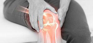
Osteoarthritis of the knee
Knee osteoarthritis is also the name of deforming arthrosis of the knee joint. The progression of osteoarthritis causes pain and further deforms the knee with possible increases in volume and inflammatory result.
In athletes the result of regular stress on the knees can develop what is known as osteoarthritis that affects the knee joint, pain is felt in the front of the affected knee, the kneecap has sharp edges). For pain followed by the crunching and clicking of the joint.
Osteoarthritis of the hip joint
Also be called osteoarthritis. This kind of arthritis severe and progresses faster than other types of this disease that leads to disability. Lesion in one hip (unilateral arthrosis) can lead to a defeat for the other (unilateral osteoarthritis). With a high probability, run existing arthrosis in the hip joints will cause damage to the knee joint and spine. There is a lameness, shortening of the affected limb, the rustling of atrophy of the thigh muscles.
uncovertebral arthrosis
When uncovertebra osteoarthritis suffers neck joint.
In addition to the above factors relate to the emergence of diseases of the joints, osteoarthritis causes uncovertebra can become a load on the cervical spine, osteochondrosis (degenerative changes of vertebrae, intervertebral discs and cartilage of adjacent tissues), disc herniation (protrusion, bulging disc of the spine).
Osteoarthritis of the neck joint is accompanied by a decrease of cartilage, compaction and deformation processes of the cervical, decrease of space, growth of bone tissue in the hand in consequence of the narrow space in the cervical region, compression of the nerve roots and narrowing of the spinal canal.
For uncovertebral arthrosis characteristic symptoms headache, neck pain, dizziness, vomiting, low sensitivity neck or feelings of tingling in this area, the increase in blood pressure.
Arthrosis of the jaw joint (TMJ)
TMJ – temporo-mandibular joint. The joint is closer to Wuhan, between the jaw and the head on both sides of the skull. The cartilage of the jaw joint not so strong as in other connection associations, so his defeat causes severe pain. The disease is common among people aged 50 years ill mostly women. The result of arthrosis of TMJ is difficulty when chewing food with pain, hearing loss, pain when talking. In this case, the causes of osteoarthritis are: a change in the bite, congenital anomalies of the jaw, jaw injury, stress, unsuccessful treatment of teeth, damaged teeth, old age. By moving the jaw, is published click and crunch when you try to open your mouth the jaw may shift to the side.
Arthrosis of the elbow joint
This type of arthritis is less common other types of disease, as the load on the elbow joint is almost negligible. If there is inflammation of the localization of osteoarthritis is swollen and red, there is a click, creak, crunch with arm movement, pain, slightly increased body temperature.
Arthrosis of the foot
This type of arthritis also be called coxarthrosis. Diseases of the ankle joint is characterized by increased fatigue while walking, edema (when connected inflammation in the process), pain (especially at night). Walking people feel the clicks in the ankle joint. In coxarthrosis injuries affect the joints, muscles, tendons, nerves and blood vessels. The weakening of the tendons causes subluxation (the joint surfaces are partially in contact). When dislocation of the articular end are completely inconsistent.
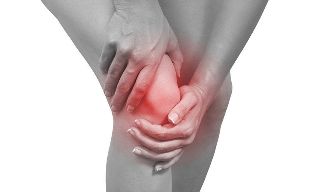
The symptoms of osteoarthritis
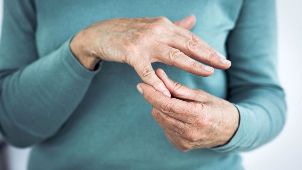
In the early stages of arthritis symptoms may not bother, but then the disease enters its momentum, and develop the first and subsequent signs. The main symptom of osteoarthritis is pain depending on the site of the damage (in the jaw and neck, shoulders, spine, elbows, wrists, fingers and feet, knees, hips and ankle joint). Pains are dull in nature, but with the heavy course of the disease will increase and become unbearable. The main symptoms of osteoarthritis:
- starting pain (joint stiffness after sleep or rest, held during the day);
- mechanical pain (occurs after employment, exertion, running, exercises, walks);
- night pain (venous stasis provokes pain at night);
- weather dependent (changes wait worsen the condition);
- crunching, creaking, crackling or uncharacteristic, clicking in the joint;
- limited mobility;
- deformity of the affected joints;
- cramps, unpleasant muscle spasms;
- lameness;
- redness and swelling at the site of the affected joint (additional inflammation).
Diagnosis of osteoarthritis
Self-diagnosis of "arthritis" do not deliver. With a disturbing pain, crunching in the joint should be treated first to the therapist. He will examine the patient and give a direction to the right doctor, orthopedist, traumatologist, a rheumatologist, a surgeon or neurologist.
Arthritis diagnose a podiatrist. Before examination, the doctor collects the anamnesis (information about the patient, hereditary or chronic disease), and doing a review. The patient is then sent for examination.
Laboratory examination
In the diagnosis of osteoarthritis may need the following tests:
- General analysis of blood (blood from finger);
- biochemical analysis of blood (rheumatology test, taking blood from veins);
- urine;
- analysis of synovial fluid or puncture of the patient's joint (the fence by means of the injection into the joint).
Instrumental diagnostics
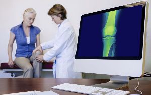
X – ray- this study will show the stage of arthrosis, the slightest changes in the structure of the joint and deformation using x-rays. Before the examination the patient has to remove jewelry so as not to distort the result in the picture. You need to remove the clothes from that part of the body that will be examined (if you need an x-ray of the knee, you need to remove the pants; the gypsum is not removed). Then the doctor says what position you need to take to images of the patient area. At the time of diagnosis the patient is served a lead apron toward the groin, and the area of the thyroid gland, the apron will prevent passage to these places x-ray radiation. Then the picture is taken.
The x-ray beam to pass through meters of fabric, but is absorbed by the solids (bones, teeth). The soft tissue on x-ray is obtained of a dark color, and solid fabric light. X-ray is the best diagnostic method of bone tissue, as the photographs clearly reflected bones and joints.
CT (computed tomography) – a method of radiological diagnosis, shows the organ, bone or tissue in the incision in layers. A CT scan is performed if x-rays are insufficiently informative and the arthritis cannot be determined. The procedure is performed in a few minutes. The patient lies on a table after removing clothing and metal jewelry. Then a ring of detectors (scanner) rotates around the reclining patient, and snapshots. Lying during the CT scan have still.
MRI (magnetic resonance imaging) – a method of radiological diagnosis. Different from CT because it is better to distinguish m of fabric, but does not distinguish between the presence of calcium in the bones, unlike a CT scan that examines bone tissue. MRI in osteoarthritis assigned to learn more about the condition of the surrounding tissues or to investigate the cause of the disease. Diagnosis using MRI is expensive, but informative.
Sonography (ultrasound) is a safe non-invasive method, without radiation and harmful substances. The doctor led sensor at the problem area and the screen shows the internal structure of the joint. This method of diagnosis of osteoarthritis of affordable, safe and informative.
Forecast
Arthritis has a good prognosis when it comes to the life of the patient, the disease is not fatal, but complications can lead to disability. However, to completely cure the arthrosis is impossible, since the age of disruption is faster than the recovery of damaged areas. The disease progresses slowly and can develop over decades. Finding the first signs of osteoarthritis can slow down further deformity and dysfunction of the joint with treatment at the initial stage.
Prevention
Often resulting in uneven, unnecessary stress suffer knee and hip joints, so you need to calculate power load and get rid of excess weight, if any to make the joints work properly and smoothly.