


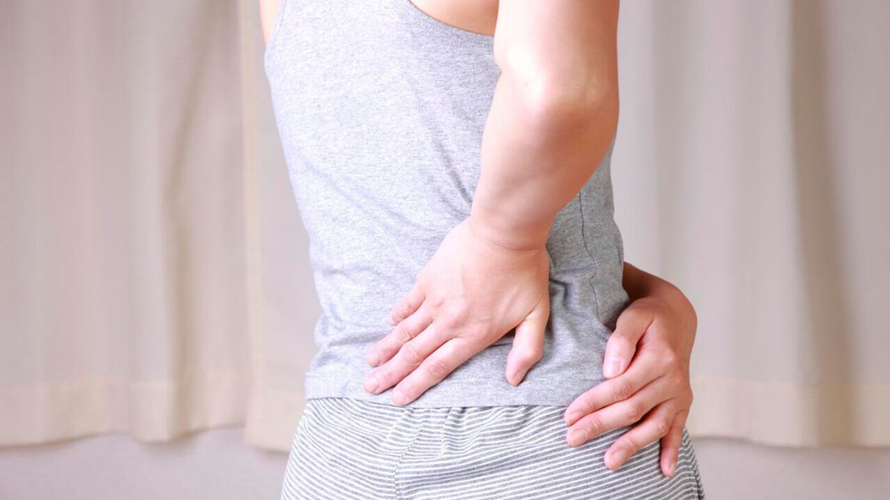
One of the frequent symptoms with which patients come to the medical facility is pain in the hip joint. The causes, treatment and possible diseases that cause such a manifestation cannot be identified without qualified medical care. Discomfort in any part of the musculoskeletal system can indicate the development of serious pathologies, so hip joint dysfunction should not be ignored.
Anatomy of the hip joint area
The hip joint plays an important role in motor activity. This is one of the largest human joints, which can withstand heavy loads in a standing position, as well as when walking upright.
Bones that form a joint
The hip joint is formed by the head of the femur and the acetabulum of the unnamed pelvic bone - the most powerful and largest parts of the human skeleton. The minimum number of anatomical elements of the joint ensures its strength and reliability, the ability to withstand body weight during movement. Most pathologies of the hip joint begin with damage to the acetabulum, the immovable part of the joint. It is shaped like a pelvis, the center of which is directed upwards at a slight angle, which ensures an even distribution of the load between the pelvic bones.
The glenoid cavity is a strong and solid formation, which consists of 3 types of pelvic bones:
- ileal
- ischial
- shy
The most vulnerable area of the joint cavity is in children whose bone tissue is not strong enough. Due to the presence of a small bone ridge on the edge of the depression, the head of the femur is completely immersed in the "bowl", which provides strong support to the extremity. The moving part of the joint is the femur (head, neck, greater and lesser trochanters). The shape of the head corresponds to the cavity of the joint cavity. It is coveredcartilaginous tissue, which ensures perfect alignment of the joint elements and their smooth sliding. In the middle of the head is a strong ligament that connects the bone to the acetabulum, providing additional grip and support.
The neck emerges from the head of the femur at an obtuse angle, which ensures the mobility of the joint and the even distribution of the load between the limbs. Trochanters are bony projections to which muscle tendons are attached.
Fabrics and structures
The normal functioning of the joint is ensured by various structures, each of which performs the appropriate functions.
Blood supply, performance and reduced joint sensitivity ensure:
- Ligaments and tendons surround the joint from all sides, covering and protecting the femur and its neck, as well as the socket itself.
- Cartilage covers the head of the femur and part of the acetabulum.
- Subcartilaginous areas are bony tissue consisting of cells and connective extracellular substance.
- The joint membrane or capsule is the source of a special secretion - synovial fluid for lubricating the parts of the joint.
- The acetabular labrum connects the edge of the acetabulum and the transverse ligament.
The hip joint is supplied with nutrients through a fairly isolated network of vessels and arteries. The blood supply to the internal parts of the joint is provided by the acetabular branch of the sealing artery, while the capsules, ligaments and surrounding muscles are supplied through the deep arteries of the upper leg and buttocks.
Anatomical formations located next to the hip joint
Often the cause of pain in the hip joint is damage to the anatomical structures located next to it. These elements include:
- Skin and subcutaneous tissue - the outer covering of the body
- The muscles of the thighs, pelvis, lower back and buttocks ensure the mobility of the joint and additionally strengthen it from the outside
- Extra-articular ligaments - perform a strengthening function, they are located around the joint capsule
- Periarticular bursae are bundles of connective tissue that prevent friction between soft and hard tissues
Risk factors
Inflammatory processes in the pelvic area are caused by mechanical damage or damage by certain types of bacteria. In this case, both the joint elements and the anatomical formations surrounding them may be exposed to pathological effects.
As a rule, one or more structures are ignited:
- skin
- muscles
- ligaments (extra-articular, femoral heads)
- periarticular bursa
- TBS capsule
- cartilage
- acetabular labrum
- subcartilaginous areas
Pain in the hip joint is often caused by harmful microorganisms that cause the development of infectious arthritis. Other reasons are also common:
- disorders of the immune system
- joint injuries due to excessive physical activity
- old age
- metabolic disorders
- other diseases
Characteristics of pain
When diagnosing hip pain, additional symptoms play an important role, which may indicate the root cause of the problem.
Pain in the hip joint and spreads to the leg
If the pain from the painful joint radiates to the groin, knees or buttocks, then the problem is most likely caused by damage to the nerve that innervates the leg area due to one of the following reasons:
- joint tumor
- infectious arthritis - occurs due to damage by pathogens
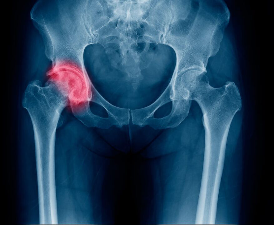
- femur fracture (in the head or neck area)
- Legg-Calvé-Perthes pathology - necrosis of the cartilage tissue of the femoral head
- juvenile epiphysiolysis - disorder of the structure of the joint head and its inflammation
Pain in the hip joint, radiating to the leg, can signal pathologies of the cartilage tissue and periarticular structures, lack of lubrication of the joint and damage to the synovial membrane. Painful symptoms may appear suddenly or increase gradually.
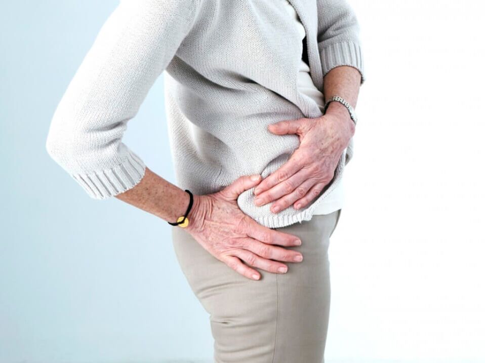
Pain when walking
Pain in the hip joint can occur during walking if the acetabulum comes into contact with the cartilaginous tissue of the femoral head, resulting in an inflammatory process. The cause of this phenomenon can be mechanical damage, inflammation of the anatomical formations located next to the joint.
Based on the intensity of pain in the hip joint when walking, you can identify the root cause of the problem:
- discomfort that occurs at the beginning of walking, gradually decreasing - a sign of inflammation of the periarticular bursa
- discomfort that gradually increases from the moment you start walking - inflammation of the articular surfaces of the hip joint
- continuous pain of high intensity, accompanied by impaired functionality of the joint - occurs in dislocations and fractures
- the pain occurs closer to night - a consequence of the deformation of the cartilage of the head of the femur and (or) the acetabulum, which rub against each other and become inflamed
- pain of moderate intensity is a sign of minor injuries and bruises
Pain when abducting the leg
Pain during leg abduction is caused by inflammation of tissues and structures that enable movement: muscles, periarticular bursae, tendons. Similar symptoms are often the result of myositis (inflammation of muscle tissue), bursitis (inflammation of the periarticular bursa) and tendinitis (inflammation of tendons).
Causes
In most cases, pain in the pelvic area is caused by a patient who has one of the following problems:
- arthritis
- coxarthrosis
- trochanteric bursa bursitis
- tendinitis
- infectious pathologies
- hereditary diseases
- tumor formation in the pelvic region
Without timely treatment, each of these causes can lead to severe complications, including loss of joint mobility.
Arthritis
Arthritis (coxitis) is a disease of joint tissue caused by disorders of the immune system or damage caused by pathogens: viruses and bacteria.
Symptoms of arthritis:
- temperature increase
- pain and swelling in the joint area
- motor impairment
The disease occurs in acute, subacute and chronic forms.
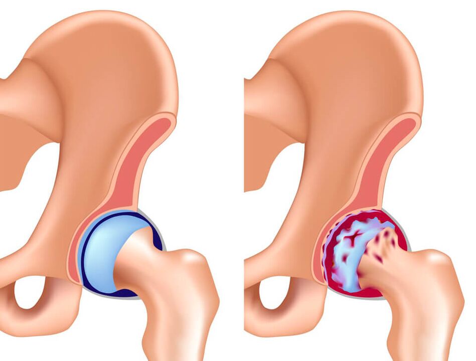
Coxarthrosis
Another name for coxarthrosis is osteoarthritis of the hip joint. This pathology is caused by metabolic disorders in cartilage tissue, which results in their death. The cause of this phenomenon can be injuries, disturbed blood supply, excessive physical activity, age over 45 and heredity. The main symptom of coxarthrosis is pain in the lower back, groin and buttocks, which gradually increases during physical activity and leads to lameness. Discomfort decreases during periods of inactivity.
Bursitis of the trochanteric bursa
The presence of an inflammatory process in the bursa (trochanteric bursa) is characterized by the appearance of intense pain in the joint area. Athletes and elderly people are susceptible to this disease. The main symptom of bursitis of the trochanteric bursa is pain in the area of the greater trochanter, which intensifies when trying to lean on the affected leg.
Tendinitis
Inflammation of tendons is called tendinitis. This is a disease that occurs in acute or chronic forms and leads to degenerative changes in the tissue. Often, the pathology occurs in athletes who do not follow the running technique, as well as after heavy loading of the hip muscles.
Tendinitis is usually a complication of another disease:
- pathology of the thyroid gland
- metabolic disorders
- arthritis
- arthrosis
- inflammatory process of systemic or infectious origin
- hip dysplasia
Tendonitis causes the patient discomfort during movement, pain, changes in gait and clicking when walking in the joint area.
Infections
Some infectious diseases cause inflammation of joint tissue, as well as nearby anatomical structures, resulting in intense pain in the hip joint. Most often, the following pathologies have similar symptoms:
- Aseptic necrosis of the femoral head is a disorder of the blood supply to the groin, resulting in tissue death. The pain associated with this disease is acute and intense. The problem is more common in men.
- Purulent arthritis is a serious disease that requires immediate treatment. If you don't seek immediate medical attention, sepsis can occur. Associated symptoms are general intoxication, pain and swelling in the area of the affected joint, difficulties in motor activity.
- Tuberculous arthritis is common in children and is characterized by slow progression. Associated symptoms are increased fatigue, decreased motor activity and muscle atrophy. Pain of varying intensity increases when a purulent abscess appears.
Infectious pathologies of the hip joint lead to severe complications and therefore require urgent treatment.
Hereditary diseases
Hereditary pathologies of the hip joint usually occur between the ages of 1 and 10, and are characterized by pathological changes in the tissue of the glenoid cavity and/or the femoral head. The most common hereditary disease affecting the hip joint is Legg-Calvé-Perthes syndrome, which is characterized by pain and gait disturbance due to the death of cartilage tissue in the joint.
Bone and soft tissue tumors
Benign or malignant bone and soft tissue growths in the hip joint can cause pain when walking or resting. The tumor can occur in bone tissue (osteomyelitis), cartilage tissue (chondroblastoma, chondroma), osteochondral tissue (osteochondroma). As a rule, neoplasms cause discomfort and are felt during palpation. Benign tumors are treated surgically, and some of them can transform into cancerous tumors.
Soft tissue tumors of the thigh:
- lipoma
- rhabdomyoma
- fibroma
- hemangioma
- neuroma
An oncologist is involved in the diagnosis and treatment of tumors in the hips and pelvis.
What to do
With severe pathologies of the hip joint, a person feels severe pain. Discomfort in the pelvic area is a reason to visit a medical facility for examination and treatment.
Special attention should be paid to the intensity of the pain:
- Lungs- appear with bruises after an injury. Cold should be applied to the painful area to reduce swelling. To reduce pain, it is recommended to take non-steroidal anti-inflammatory drugs. It is recommended to consult a doctor.
- Moderately- they usually occur in diseases of the hip joint, accompanied by impaired motor activity and elevated body temperature. Discomfort increases during physical activity. Consultation with a rheumatologist is necessary.
- Strong- occur due to dislocations and fractures. Accompanied by limitation or impossibility of physical activity. In cases of severe pain in the hip joint caused by an injury, it is necessary to contact emergency services.
There are many folk recipes that are used for pain in the hip joint. It is important to remember that all of them are suitable for symptomatic treatment and pain reduction, but will not help to eliminate the cause of the problem. Effective treatment is carried out exclusively under the supervision of a qualified physician.
Which doctor should I contact?
If you have hip pain, contact your family doctor or general practitioner, who will refer you to a specialist. Diseases of the musculoskeletal system are treated:
- traumatologist— pain in the hip joint due to physical activity, sprains, falls and other injuries
- rheumatologist- sudden onset of joint pain for no apparent reason
You may also need to consult with other doctors: surgeon, oncologist, infectious disease specialist, etc.
Diagnostics
The first important stage in diagnosing pain in the hip joint is an external examination, which necessarily includes taking an anamnesis and palpation. Depending on the severity of the disease and the patient's complaints, laboratory tests and instrumental diagnostic methods are prescribed:
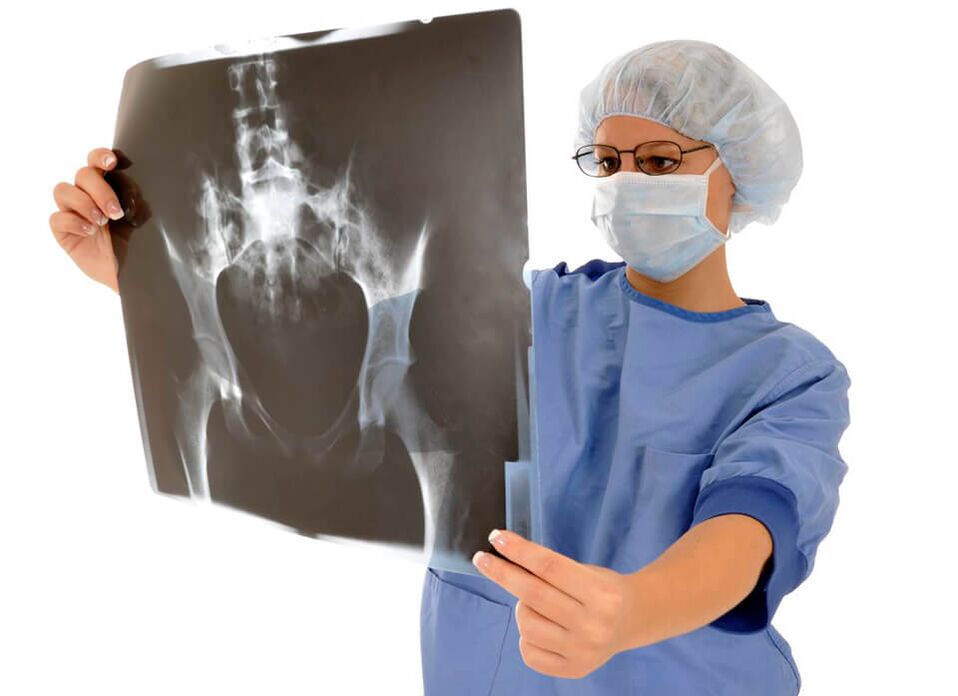
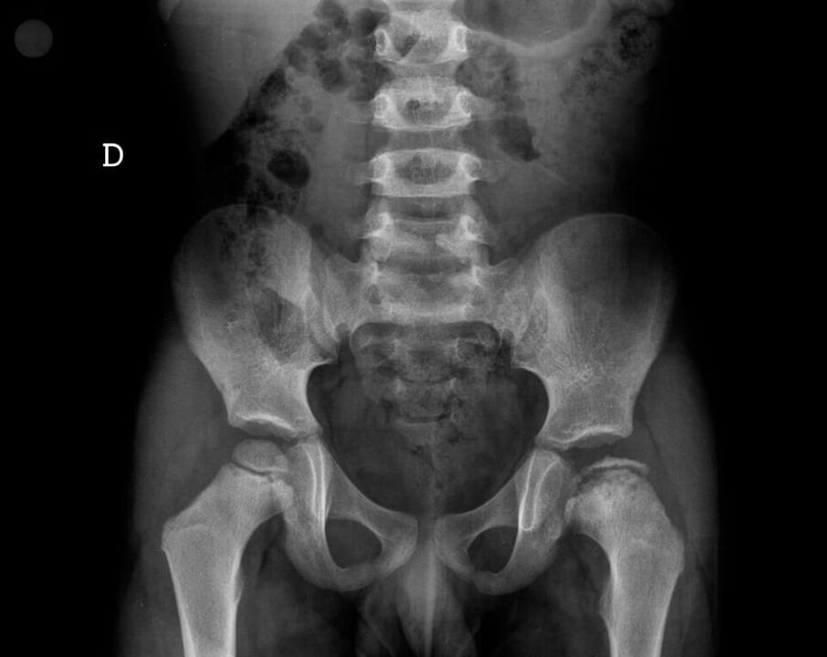
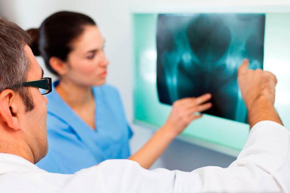
- radiography- the use of X-rays to examine a certain area of the body
- CT and MRI- modern precise diagnostic methods that allow you to get highly informative images of the joint and the area around it
- microbiological examination of a sample of biological materialenable detection of the presence of pathogenic microorganisms: viruses and bacteria
- immunological blood test- allows you to identify immune disorders, determine the presence of certain autoantibodies
- arthroscopy (endoscopic examination)— examination with a probe, the possibility of taking a sample of joint tissue for further research
- laboratory examination of effusion- taking a sample of intra-articular fluid during puncture and determining the causative agent of an infectious disease in it, checking sterility
The combination of several diagnostic methods allows us to identify the cause of pain in the hip joint with great precision.
Treatment

Treatment of pain in the hip joint should be prescribed by a doctor based on examination and diagnosis. As a rule, drug therapy or surgery is prescribed.
Medicines
Treatment of pain in the hip joint should be comprehensive, aimed at eliminating the symptoms, and most importantly, eliminating the cause of the problem. For this purpose, drug therapy is used, which includes the use of:
- nonsteroidal anti-inflammatory drugs- they help reduce pain, relieve swelling
- means for improving microcirculation- they help restore blood circulation and nutrition of joint tissues
- chondroprotectors- stimulates the renewal of cartilage tissue
- muscle relaxants- reduce pain, improve blood flow in the damaged area
- hormonal drugs- to relieve pain and suppress inflammation
In the treatment of pain in the hip joint, physiological procedures are very effective: massage, acupuncture, cryo- and laser therapy. Special therapeutic exercises and manual therapy are also used.
Surgical
Surgical intervention is indicated in advanced cases when conservative treatment does not help the patient. This includes partial or complete replacement of the diseased joint with a prosthesis.
Prevention
Reducing the load on the legs will slow down the pathological processes inside the joint, so obese people are recommended to start losing weight.
Preventive measures will help reduce pain in the hip joint:
- regular walking
- physiotherapy
- a balanced diet rich in vitamins A, C, E
Timely consultation with a doctor in the first stages of the disease increases the effectiveness of treatment, and also reduces the risk of complications and serious consequences for the body.